|
As the chief technician at the Electron Microscopy Unit, Research Branch, Agriculture and Agri-Food Canada in Ottawa, Mr. Ann-Fook Yang is responsible for smooth operations of the laboratory. He takes care of several electron microscopes and all ancillary equipment and ensures that the microscopes are available to agricultural scientists and technicians in disciplines as diverse as entomology, plant science, and biotechnology. In addition, he also teaches electron microscopy techniques to other scientists' technicians. Such procedures may include the preparation of samples for conventional scanning electron microscopy as well as immunogold labeling of various structures for transmission electron microscopy.
(Environmental SEM)
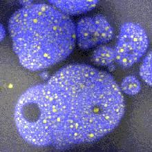
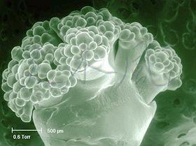
Two years ago, the laboratory acquired a new environmental scanning electron microscope for work on hydrated specimens in plant science, entomology, and elsewhere. When checking how such a microscope would produce sharp images of fresh samples, Mr. Yang examined also specimens which may be characterized as foods or closely related to foods. Vegetables such as cauliflower and broccoli are excellent examples where environmental scanning electron microscopy (ESEM) proves useful in visualizing the developing of the miniature florets as shown below.
Environmental SEM (ESEM) was done using small specimens (less than 4 mm in diameter) placed directly in the well of the cooled microscope stage maintained at +1°C and a pressure of 0.6 Torr in the chamber. The cut surface of each specimen was sealed with colloidal graphite to prevent water loss.
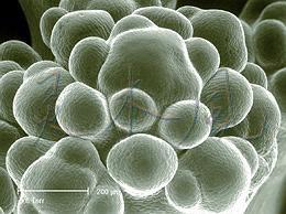
The image of cauliflower at left was obtained at 0.6 Torr and 1°C. The bar indicates 500 µm. Details of the developing florets are shown at a higher magnification at right (bar = 200 µm). There are many macroscopic images of cauliflower available on the Internet, for example as an illustration to the description of this vegetable also providing advice on growing. The internal structure of a cauliflower head is an example of fractals.
Two micrographs of broccoli, another vegetable of the Brassica genus, are shown above. The bar at left is 1 mm long and the bar at right is 500 µm long;. Broccoli is an example of another kind of fractal. Information on various aspects of growing broccoli may be found on the site of the University of Illinois.
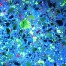
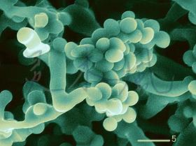
The images of cauliflower and broccoli were obtained at relatively low magnifications. However, ESEM may also be used at considerably higher magnifications to study another group of organisms such as fungi. When fungi, commonly called "moulds", appear on foods, they are the sign of spoilage to most North American and European consumers. Aspergillus, Sclerotium, Rhizopus stolonifer (the bread mould), Fusarium, and Penicillium ssp. are only a few of food-spoiling fungi. A characteristic odour which develops in citrus fruits infected by Penicillium italicum or P. digitatum is known to most consumers who have left a lemon, a grapefruit, or an orange in the refrigerator for too long. Transfer of a small piece of the Penicillium digitatum fungus into an ESEM and examination at high humidity and low temperature inside the microscope makes it possible to quickly obtain images of the hydrated hyphae and spores without any additional preparatory steps (green image below at left).
Fungi (moulds) are not common in North American foods and are mostly limited to Camembert and Blue (mould-ripened) cheeses made with Penicillium camemberti and Penicillium roqueforti, respectively. However, since yeasts such as Saccharomyces cerevisiae also belong to fungi, leavened bakery products such as bread may also be included in the list of foods containing fungi.
In Asian foods, fungi are considerably more common as part of the foods. For example, furu is a traditional Chinese food which has a foie gras-like texture and salty taste. It is made by fermenting soy protein curd (tofu) with Actinomucor elegans, Mucor hiemalis, Mucor silvaticus, Rhizopus chinensis and similar fungi. Monascus purpureus, Monascus anka and similar fungi are used to provide pleasing reddish colouring. Furu is also consumed in Japan.
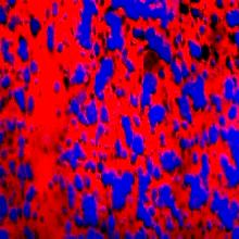
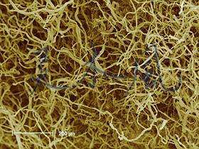
The thick mat of fungal hyphae (the brown image above at right) was obtained by conventional SEM, the first of the two alternative procedures listed below:
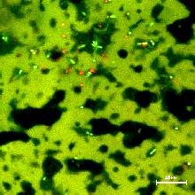
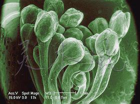
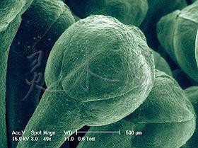
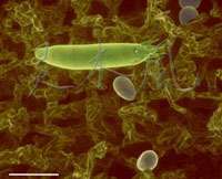
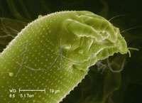
ESEM was also used to examine what at the first glance appeared as rust on pear leaves. The reddish brown spots, several mm in diameter, are also known to appear as defects on the surface of pears. The pest causing the damage, however, is not a fungus but the pear leaf blister mite. Lifting the blister gently under a dissecting microscope reveals minute mites foraging on the leaf tissue, and also unhatched eggs (top image at left). At a higher magnification, ESEM shows details of the mite's front end (bottom image at left).
Although the micrographs shown do not strictly show the internal microstructure of foods, they demonstrate the potential which this kind of microscopy has for agricultural research, foods including. The original black and white micrographs, obtained in digital form, have been coloured.
©A.-F. Yang 2000
|
|
Mark Auty, Ph.D.Mark Auty is a food microscopist with 34 years of experience and is currently a Research Officer at the Dairy Products Research Centre, Moorepark, (a division of Teagasc Co. Cork, Ireland. He has studied a wide variety of foods, mainly for industrial clients, using light and electron microscopy techniques. Recently he has specialized in confocal microscopy. Confocal Laser Scanning Microscopy (CLSM) of Dairy Products
Powders
Results indicated that powders with high free fat contents (as determined by solvent extraction) did not necessarily have high levels of surface fat but had large fat droplets within the powder particle. Powders with high surface fat contents dispersed poorly in water, but were ideal for chocolate manufacture, where fat is the continuous phase.
Chocolate
Additional information about chocolate may be found at these links:
Cheddar cheese Cheddar, cream cheese, analogue Mozzarella and Mozzarella cheeses were examined using the new dual staining technique developed for powders. The distribution of protein and fat in each of the cheeses varied depending on the variety. Cheddar cheese consisted of irregular fat droplets surrounded by a protein matrix. Cream cheese consisted of aggregates of small fat droplets and protein. Analogue Mozzarella was an emulsion of rounded fat droplets in a homogeneous protein matrix containing crystals of emulsifying salts. Mozzarella consisted of elongated protein fibres containing small fat droplets with some fat droplet clusters entrapped between protein fibres.
Microbiology
Dr. Mark E. A. Auty
tel: +353 25 42442
|