 |
 |
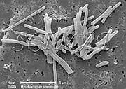
| Bar: 5 µm | Bar: 2 µm |
|
|
| Bar: 5 µm | Bar: 2 µm |
|
Mycobacteria are unicellular, aerobic, Gram-positive bacteria, taxonomically related to the antibiotic-producing streptomycetes. They are unusual among bacteria in that they have an enormously thick, hydrophobic cell wall that prevents desiccation. Many mycobacteria are harmless and useful because they degrade organic matter in soil. Better known are, however, the few pathogenic mycobacteria which cause tuberculosis (M. tuberculosis, M. africanum, M. bovis) and leprosy (M. leprae). Mycobacterium smegmatis was isolated from smegma and may also be found in the soil. The bacteria are nonmotile slender rods with branching or Y shapes. Mycobacteria protect themselves with an outer lipid bilayer, which is the thickest biological membrane hitherto known and has an exceptionally low permeability rendering mycobacteria intrinsically resistant against many antibiotics. The membrane serves as an effective permeability barrier but contains porins that serve functions analogous to those observed in Gram-negative bacteria. Electron microscopy shows the fascinating world of mycobacterial porins. Mycobacterium smegmatis, a rapidly growing mycobacterium, has only recently been reported to cause disease in humans. In the studies of tuberculosis, the rapidly growing non-pathogenic M. smegmatis is in model experiments compared to severely pathogenic M. tuberculosis and M. avium. It was also used to study the ability of the pathogenic M. tuberculosis to survive inside human macrophages. Another potential use may be similar to the use of Mycobacterium terrae as a surrogate for M. tuberculosis in disinfectant tests or in the evaluation of chemical disinfection of drinking water. |
For access to micrographs of other microorganisms please visit the home page.
Commercial interest in micrographs should be directed to photo banks such as
CMSP,
MS,
PR,
VU,
etc.
A bacterial culture was provided by Dr. Syed A. Sattar
at the Centre for Research on Environmental Microbiology (University of Ottawa).
The cells were examined by scanning electron microscopy by Milos Kalab.
| Updated: May 24, 2007.
©SCIMAT 2007 |