|
Clostridium difficile is an anaerobic, gram-positive bacterium, which may be found in marine sediment and in sand, in camel, horse and donkey dung, feces of dogs, cats and birds, human genital tract, intestinal tract of humans and in their feces and generally in hospital environment. It is responsible for nearly all gastrointestinal infections, ranging from mild diarrhea to severe or even fatal colitis, that follow antibiotic therapy.
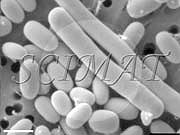
(Long bacteria may be seen in the left image obtained by scanning electron microscopy at a lower magnification. Several short spores are also noticeable scattered among the bacteria. The right image shows mostly spores with 2 long bacterial cells.) These bacteria are less frequently found in older children and in adults. This microbial pathogen is the major cause of pseudomembranous colitis and antibiotic-associated diarrhea. Pseudomembranous colitis occurs when antibiotics (esp. clindamycin and ampicillin) suppress the normal microflora, allowing C. difficile to grow and produce toxin. Two toxins have been described.
Prevention and control of Clostridium difficile is very important. Strict adherence to hand washing and a proper handling of contaminated wastes effectively suppress the bacteria. To cope with the difficulties associated with C. difficile infections, patients suffering from Clostridium difficile-induced colitis have established their support group site.
Pseudomembranous colitis caused by C. difficile and health problems caused by other food-borne microorganisms have also been described.
The Clostridium group contains more than 60 different species of bacteria. They are commonly found in soil everywhere in the world, and some species even live harmlessly in our intestines. However, some of the Clostridia cause gas gangrene, Clostridium food poisoning, and pseudomembranous colitis.
The vegetative cells and spores of Clostridium difficile were provided for scanning electron microscopy
by Dr. Syed A. Sattar and Dr. Justo Perez
of the University of Ottawa Centre for Research on Environmental Microbiology.
The bacterial suspension was treated with lysozyme to break down
remnants of the cell wall and to obtain a cleaner preparation.
Scanning electron microscopy by Milos Kalab.
For additional information on food-borne bacteria please see these
links.
|